
CT scans reveal new insights into ancient Egyptian mummies
Note: AI technology was used to generate this article's audio.
- Advanced imaging enables non-destructive examination of ancient mummies
- New medical and historical insights extracted from centuries-old remains
Advanced CT scanning technology is revealing new details inside ancient Egyptian mummies without the need to open or damage them.
Researchers at the Semmelweis Museum of Medical History in Budapest, part of the Hungarian National Museum’s collection center, are using high-resolution imaging techniques to study a group of Egyptian mummies from different periods. The goal is to examine their internal structure, mummification methods, and the health conditions of the individuals.
Researcher Ibolyka Dudás said the aim of the scans is to produce a precise view of internal anatomy and possible medical conditions, as well as to better understand ancient preservation techniques.
Museum officials noted that some of the mummies had been studied before, but newer imaging systems now provide significantly higher precision, potentially leading to new scientific findings.
According to researchers, early analyses suggest that mummification patterns may help refine the dating of some remains, especially after carbon dating produced mixed results across samples.
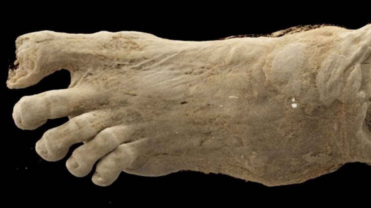
The scans have also revealed unexpected findings. One wrapped bundle previously believed to contain a bird was found to contain a human foot, while another case showed signs of osteoporosis in the individual.
Similar CT studies on mummies worldwide have identified conditions such as anemia, arthritis, and even cancer, while also helping reconstruct aspects of ancient daily life and burial practices.
Researchers say modern imaging is opening new frontiers in mummy studies by allowing internal examination without damaging artifacts that are thousands of years old.